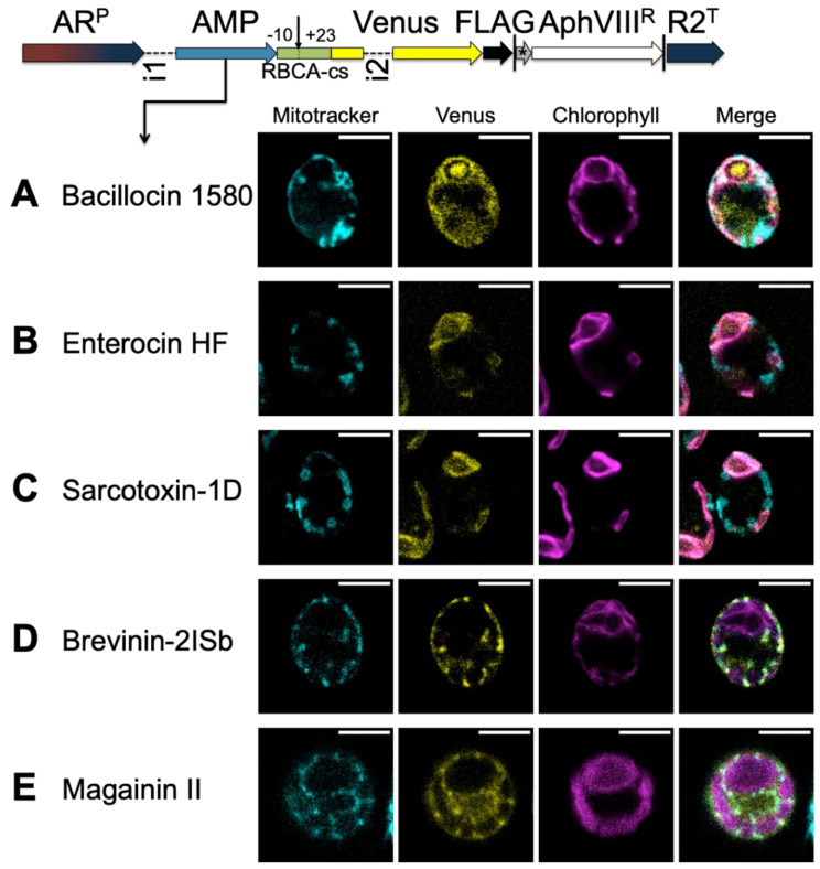
Figure 5.
AMPs function as TPs. Constructs, schematically depicted at the top of the figure, assay the targeting ability of candidate peptides fused to the Venus-FLAG reporter, driven by the chimeric HSP70-RBCS promoter and the RBCS2 5′UTR (ARP) and RBCS2 terminator (R2T), and expressed bicistronically via the STOP-TAGCAT (*) sequence with the paromomycin resistance marker (AphVIIIR). Vertical lines indicate stop codons. Expression levels in C. reinhardtii are increased by the use of introns: RBCS2 intron 1 (i1) in the 5′ UTR and RBCS2 intron 2 (i2) within the Venus coding sequence. Candidate HA-RAMPs, i.e., bacillocin 1580 (A), enterocin HF (B), sarcotoxin-1D (C), brevinin-2ISb (D) and magainin II (E) were fused to the RBCA cleavage site fragment encompassing residues −10 to +23 (RBCA-cs) and inserted upstream of Venus. The site of cleavage is indicated by a downward arrow. False-color confocal images of representative cells show mitochondria as indicated by mitotracker fluorescence in cyan, the localization of Venus in yellow and chlorophyll autofluorescence in magenta. Scale bars are 5 μm. See Figure S9 for a quantification of co-localization, Figure S10 for replicates, and Table S6 for a description of peptide sequences.

