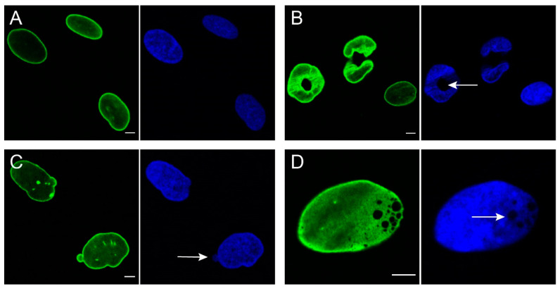
Figure 2.
Nuclear abnormalities frequently observed in human dermal fibroblasts from laminopathy patients. Green: lamin A/C staining using Jol2, Blue: DAPI staining of nuclei. (A) normal nuclei; (B) donut-like shaped nuclei with irregular lamin A/C staining. Note the hole transversing the complete nucleus (arrow); (C) nuclear blebbing: nuclear herniations with increased lamin A/C expression. Sometimes, however, lamin A/C expression can be absent in blebs (not shown). Arrow indicates presence of low amount of DNA in bleb; (D) honeycomb structures, creating an intranuclear gap (arrow). Bars represent 5 µm.

