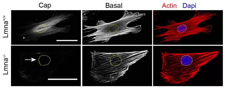
Figure 3.
Impaired directional actin cap formation in lmna−/− cells. Cells were seeded on oval microposts causing directed orientation. After staining for F-actin, confocal z-series were recorded, which show oriented actin fibers at bottom of the cell (basal) in both types of cells, along with an oriented actin cap in lmna+/+ cell. This cap on top of the cell is absent in the lmna−/− cell (arrow). Bars represent 10 µm. Adapted from Tamiello et al. [79].

