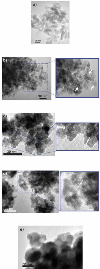
Figure 2.

Selected TEM (Transmission Electron Microscopy) micrographs of (a) P25 and of samples (b) M‐TiO2 (arrows in the inset show the occurrence of intra‐particle mesopores, deriving from the synthesis procedure depicted in Scheme 1a); (c) RM‐TiO2 (arrows in the inset show interference fringes); (d) TF‐TiO2‐200 (arrows in the inset show interference fringes) and (e) TF‐TiO2 −600.
