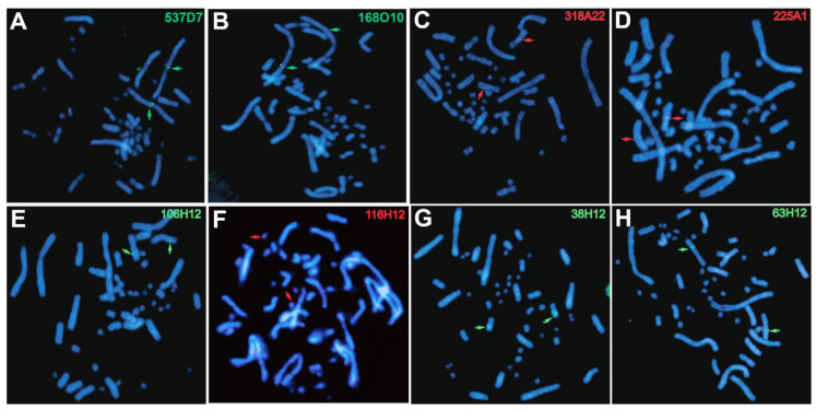
Figure 2.
Examples of chromosomal locations of BAC clones on C. picta chromosome spreads detected by fluorescent in situ hybridization (FISH) that reveal assembly errors as listed in Table 2. Top panels (A–D) show FISH results from the present study. Bottom panels (E–H) contain unpublished images showing FISH results from our previous study [12]. Green denotes probes labeled with biotin-16-dUTP and highlighted by green arrows. Red denotes probes labeled with digoxigenin-11-dUTP and highlighted by red arrows. The red hybridization signal in panel F is subtler than in other panels and zooming in may be needed for better visualization.

