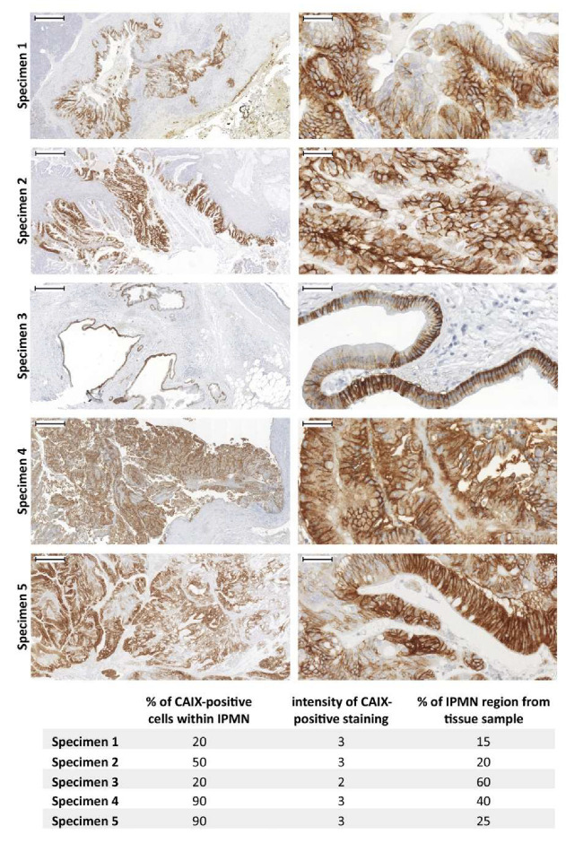
Figure 6.
Immunohistochemical staining of CAIX in IPMN lesions within pancreatic ductal adenocarcinoma (PDAC) patient samples. The set of 5 IPMN tissue specimens were immunostained using the specific anti-CAIX monoclonal antibody M75 [143] as described previously [144] (Supplementary Material). Representative pictures taken from all 5 IPMN tissue specimens (left side) were complemented with detailed pictures (right side) describing CAIX-specific staining pattern (brown). All sections were counterstained with Mayer’s hematoxylin (blue nuclei). Scale bar 500 μm (left side) and 50 μm (right side).

