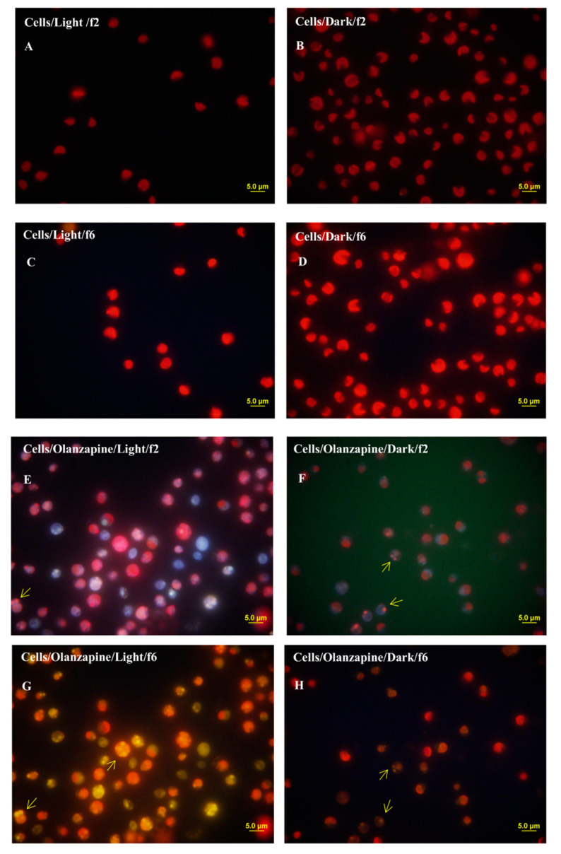
Figure 4.
Epifluorescence microscopy images of Nannochloropsis sp. cells showing the red autofluorescence of chlorophyll from chloroplasts and fluorescence of the olanzapine. Cells grown in an f/2 medium (no pharmaceuticals added to the medium) (A–D). (A,C) show cells grown autotrophically. (B,D) show cells grown in the dark (different microscope filters, f2 and f6). Cells grown in f/2 medium with OLA added to the medium (E–H). (E,G) cells grown autotrophically (microscopy filters f2 and f6). (F,H) cells grown in the dark (microscopy filters f2 and f6).

