Figure 6.
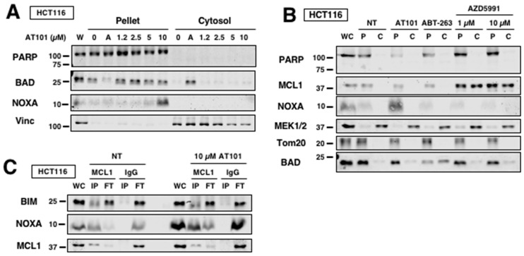
AT101 primes cells for sensitization through NOXA induction. (A) HCT116 cells were incubated with either 10 µM ABT-263 (“A”) or AT101 for 6 h and then permeabilized with digitonin and separated into pellet and cytosolic fractions. Translocation of BAD was measured by Western blotting. PARP and vinculin were used as controls for pellet and cytosol, respectively. (B) HCT116 cells were incubated with either ABT-263 (10 µM), AT101 (10 µM) or AZD5991 (1 or 10 µM) for 6 h and then permeabilized with digitonin and separated into pellet and cytosolic fractions. PARP and MEK1/2 were used as controls for pellet and cytosol, respectively. Tom20 was used as a mitochondrial marker. (C) HCT116 cells were incubated with 10 µM AT101 for 6 h and then subjected to MCL1 immunoprecipitation. Lysates were separated into immunoprecipitate (IP) and flow-through (FT) with IgG used as a negative control. BIM, NOXA and MCL1 expression were measured by Western blotting.

