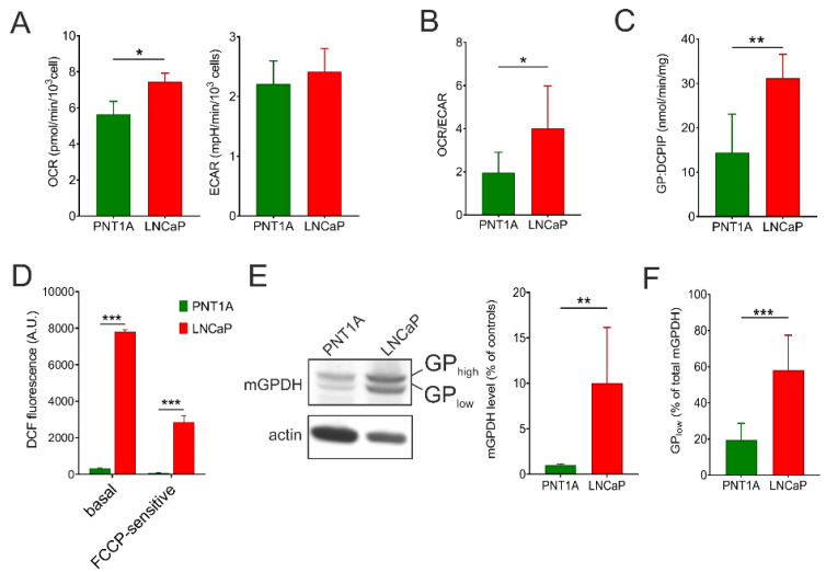
Figure 1.
Energy metabolism and mGPDH content in prostate cancer cells. The oxygen consumption rate (OCR) and extracellular acidification rate (ECAR) (A), as well as OCR/ECAR ratio (B) in prostate cancer cells (LNCaP) compared to control epithelial cell line (PNT1A). Values were calculated from the rates in basal conditions, i.e. at the presence of 10 mM glucose, and determined by a Seahorse XFe analyzer (n = 5). (C) Enzyme activity of mGPDH measured spectrophotometrically using 10 mM glycerol-3-phosphate as a substrate (n = 6). (D) ROS generation in intact LNCaP cells compared to control PNT1A measured by the CM-H2DCFDA probe. To determine the FCCP-sensitive portion of ROS production, 1 μM uncoupler was used. (E) Cell lysates (15 μg protein) were separated on SDS-PAGE and mGPDH content was analyzed by Western blotting using a specific antibody against mGPDH, actin was used as a loading control. Representative blot of 5 independent experiments is depicted. Antibody signals were quantified densitometrically as the total mGPDH levels normalized to actin levels and the results are expressed as % of control values. (F) Processing of mGPDH was determined densitometrically as a ratio of the lower band and total mGPDH content (n = 5). Data represent the means ± S.D., * p < 0.05, ** p < 0.01, *** p < 0.001.

