Figure 5.
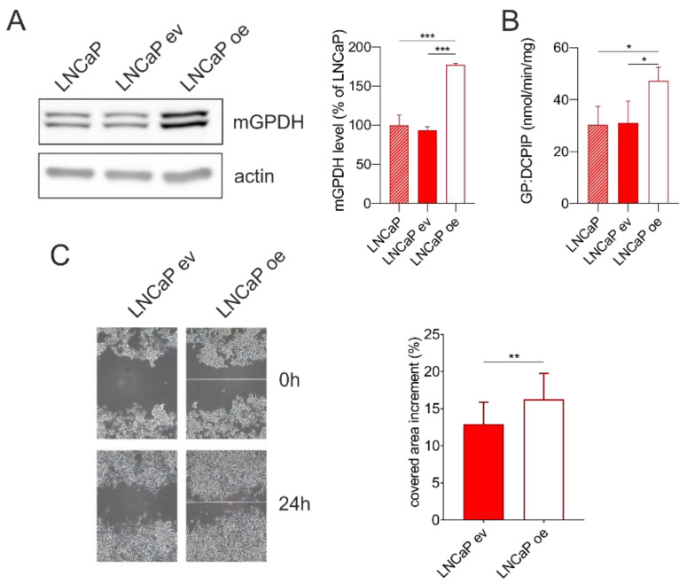
Wound healing assay in LNCaP cells without transfection, stably transfected with a control pcDNA3.1(+) vector (LNCaP ev) or vector containing mGPDH (untagged form; LNCaP oe). (A) Cell lysates (15 μg) were separated on SDS-PAGE and analyzed by Western blotting using antibodies against mGPDH and actin. A representative Western blot and quantification of total mGPDH amount is shown (n = 3). (B) Enzyme activity of mGPDH measured spectrophotometrically in cell lysates using glycerol-3-phosphate as a substrate (n = 3). (C) Analysis of cell migration by in vitro scratch assay. Images were acquired at 0 and 24 h. Representative images and quantification of covered area increment (n = 15). Data represent the means ± S.D., * p < 0.05, ** p < 0.01, ***p < 0.001.

