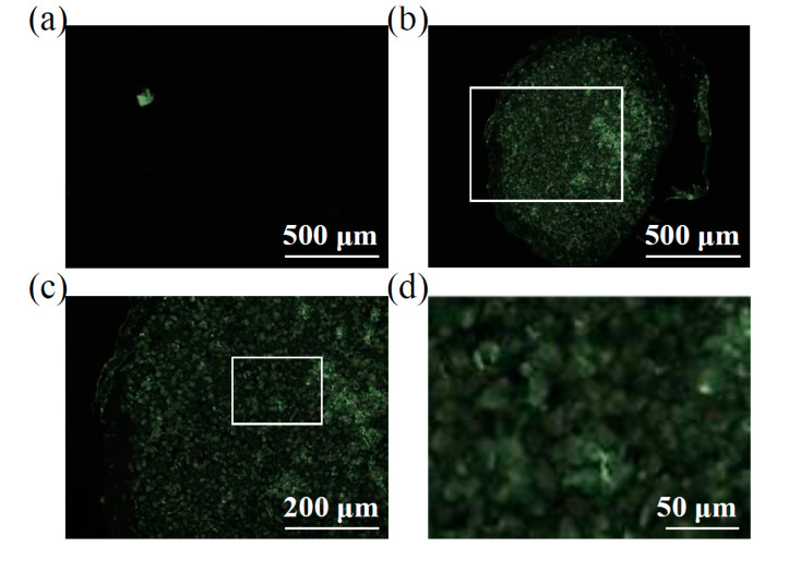
Figure 2.
ASC-EVs endpoint incorporation in chondrocyte micromasses. (a) Transversal section of a representative chondrocyte micromass not treated with CFSE-EVs showing absence of background fluorescence. A representative picture is shown. (b through d) Increasing magnification of a representative chondrocyte micromass after incubation with CFSE-EVs, with the original region of panel c depicted in panel b by the white square as well as the original region of panel d being squared in panel c. It is possible to observe a homogenous signal all over the micromass section, including the center of the pellet (b) and, with a higher magnification (c and d), fluorescence results associated with both chondrocytes and intercellular matrix. Representative pictures are shown.

