Figure 1.
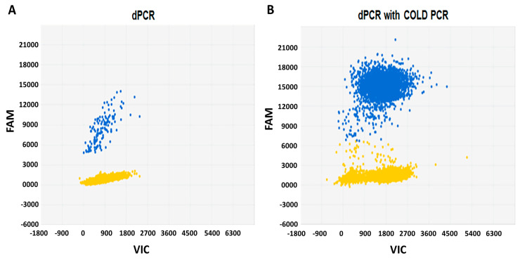
Detection of BRAFV600E by microfluidic digital PCR alone or in combination with COLD-PCR. Each panel represents a single experiment whereby DNA from BCPAP cells was segregated into individual wells and assessed for the presence of the mutant alleles using two different fluorophores (FAM and VIC). The signals from the FAM (blue) and VIC (red) dyes are plotted on the Y axis and the X axis, respectively. The yellow cluster represents the unamplified wells (negative calls). (A) Two-dimensional plots of microfluidic digital PCR reads out of 1 ng DNA extracted from BCPAP cells. Blue cluster represents the wells that were positive for the BRAFV600E mutation. (B) Results of dPCR analysis after the enrichment of BRAFV600E by COLD-PCR demonstrating 100-fold increase in mutant alleles (blue cluster). dPCR, digital Polymerase chain reaction; COLD, co-amplification at lower denaturation temperature; PCR, Polymerase chain reaction; FAM, fluorescein; VIC, 2′-chloro-7′phenyl-1,4-dichloro-6-carboxy-fluorescein.

