Figure 1.
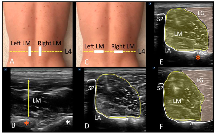
Ultrasonographic evaluation of the lumbar region. (A) Probe location for longitudinal assessment of lumbar multifidus at L4 vertebral level (yellow dotted line). (B) Longitudinal scanning view of LM (yellow arrow), and L4/5 (orange asterisk) and L5/S1 (white asterisk) facet joints visualization. (C) Probe location for transverse scanning of lumbar multifidus at L4 vertebral level (yellow dotted line). (D) Transverse scanning view of the lumbar multifidus, and visualization of the L4 spinous process (SP), the laminae (LA) and L4/5 facet joint as measurement landmark, and CSA of LM; (E), CSA of lumbar multifidus at resting (LM) and borders delimitation with lumbar longissimus muscle (LG), as well as the white enhancement of the L4 spinous process (SP), the laminae (LA), and facet joint of L4/5 (orange asterisk). (F) CSA of lumbar multifidus (LM) during muscle contraction (CAL test) and borders delimitation with lumbar longissimus muscle (LG). Abbreviations: CAL, contralateral arm lift test; CSA, cross-sectional area; LA, laminae; LG, lumbar longissimus muscle; LM, lumbar multifidus; SP, spinous process.

