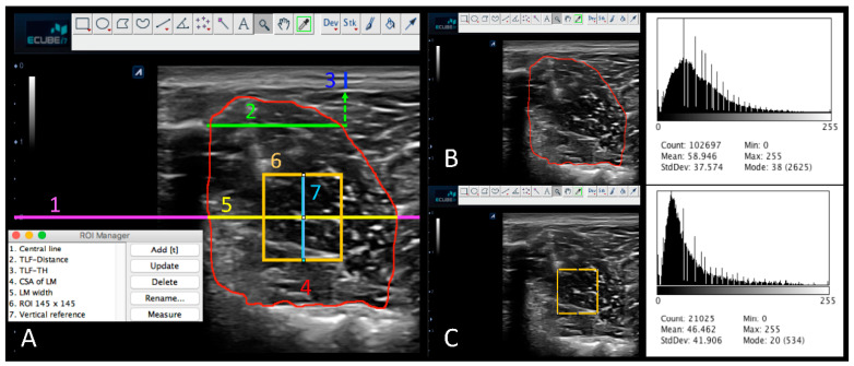
Figure 2.
ImageJ measurements of echostructure and first-order descriptors of the lumbar multifidus (LM) and the thoracolumbar fasciae (TLF). (A) Representation of the measurement protocol using ImageJ: Central line (1. Central line, magenta), Thoracolumbar fasciae distance reference (2. TLF-Distance, green), thickness (TH) of the thoracolumbar fasciae (3. TLF-TH, blue), cross-sectional area (CSA) of the lumbar multifidus at resting (4. CSA of LM red), width of the lumbar multifidus (5. LM width, yellow), region of interest (ROI) of lumbar multifidus at midpoint (6. ROI 145 × 145 pixels, orange), and vertical line reference for ROI adjustment (7. Vertical reference, cyan). (B) CSA of LM histogram displayed. (C) ROI of LM histogram displayed.

