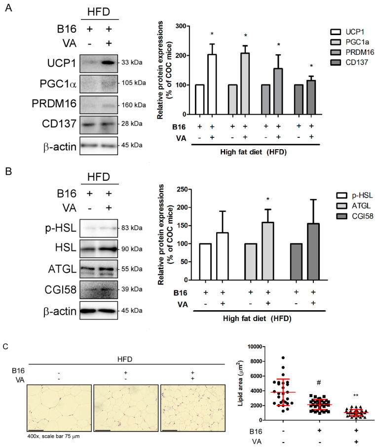
Figure 3.
Effect of VA on iWAT of COC mice. (A) Protein levels of UCP1, PGC1α, PRDM16, and CD137 were analyzed by a Western blot analysis. Results were expressed relative to β-actin. (B) Protein levels of p-HSL, ATGL, and CGI58 were analyzed by a Western blot analysis. Results were expressed relative to β-actin, except p-HSL, which was normalized to total HSL. (C) H&E staining was performed in paraffin-embedded iWAT (magnification ×400, scale bar 75 μm), and average lipid droplet size was measured. All data are expressed as mean ± SEM (n = 3); # p < 0.05 vs. HFD-fed control mice; * p < 0.05 and ** p < 0.01 vs. vehicle-treated COC mice. COC, cancer–obesity comorbidity; HFD, high fat diet; VA, vanillic acid; iWAT, inguinal white adipose tissue.

