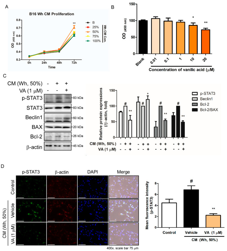
Figure 6.
Effect of VA on adipocyte CM-treated B16BL6 melanoma cells. (A) The cell growth rate of B16BL6 melanoma according to the concentration (0%, 25%, 50%, 75%, and 100%) of CM diluted in complete medium was measured by an MTS assay. (B) Cytotoxicity of VA in B16BL6 melanoma was determined by an MTS assay. (C) Protein levels of p-STAT3, Beclin-1, BAX, and Bcl-2 were analyzed by a Western blot analysis. Results were expressed relative to β-actin, except p-STAT3, which was normalized to total STAT3. (D) Expression of intracellular p-STAT3 was evaluated by an immunofluorescence staining (magnification ×400, scale bar 75 μm). All data are expressed as mean ± SEM (n = 3); # p < 0.05 vs. untreated B16BL6 melanoma cells; * p < 0.05 and ** p < 0.01 vs. CM-treated B16BL6 melanoma cells. CM (Wh), white adipocyte conditioned media; VA, vanillic acid.

