Figure 1.
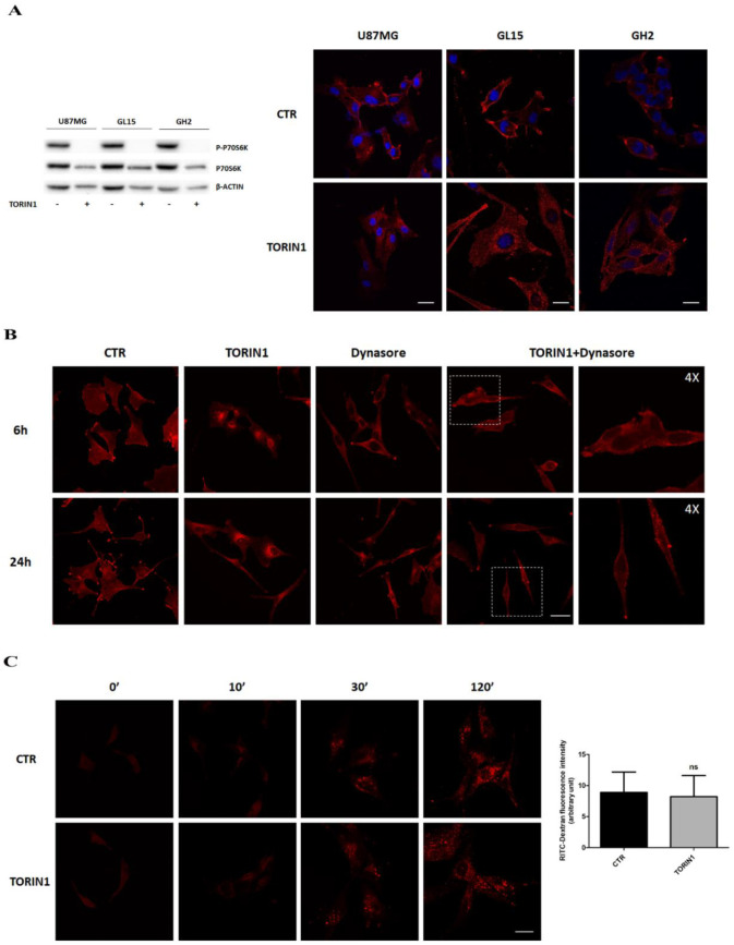
Epidermal Growth Factor receptor (EGFR) internalises into Glioblastoma multiforme (GBM) cells upon mTOR inhibition. (A) U87MG (upper panels), GL15 (middle panels) and primary GH2 cells (lower panels) were cultured in complete medium (DMEM) (CTR) or in DMEM in the presence of 250 nM Torin1 for 18 h. Immunocytochemistry and confocal analysis for EGFR localisation (red) were then performed. Hoechst 33342 was used to stain nuclei (blue). Scale bar, 30 μM. Western blot analysis of P-p70S6K and p70S6K was performed to check mTOR pathway inhibition by Torin1. β-actin was used as loading control. The blots are representative of three independent experiments. (B) Immunocytochemistry and confocal analysis for EGFR localisation (red) were performed in U87MG cells, upon 6 h and 24 h Torin1 treatment in the presence or absence of 100 μM Dynasore. Scale bar, 30 μM. A 4× magnification is shown for the right panels representing cells treated with Torin1 plus Dynasore. (C) U87MG cells were incubated with 0.5 mg/mL Rhodamine Dextran for the indicated times and its uptake within the cells analysed by image capturing at the confocal microscope. Scale bar, 30 μM. Fluorescence quantification of Dextran uptake at 120′ is shown in the right panel.

