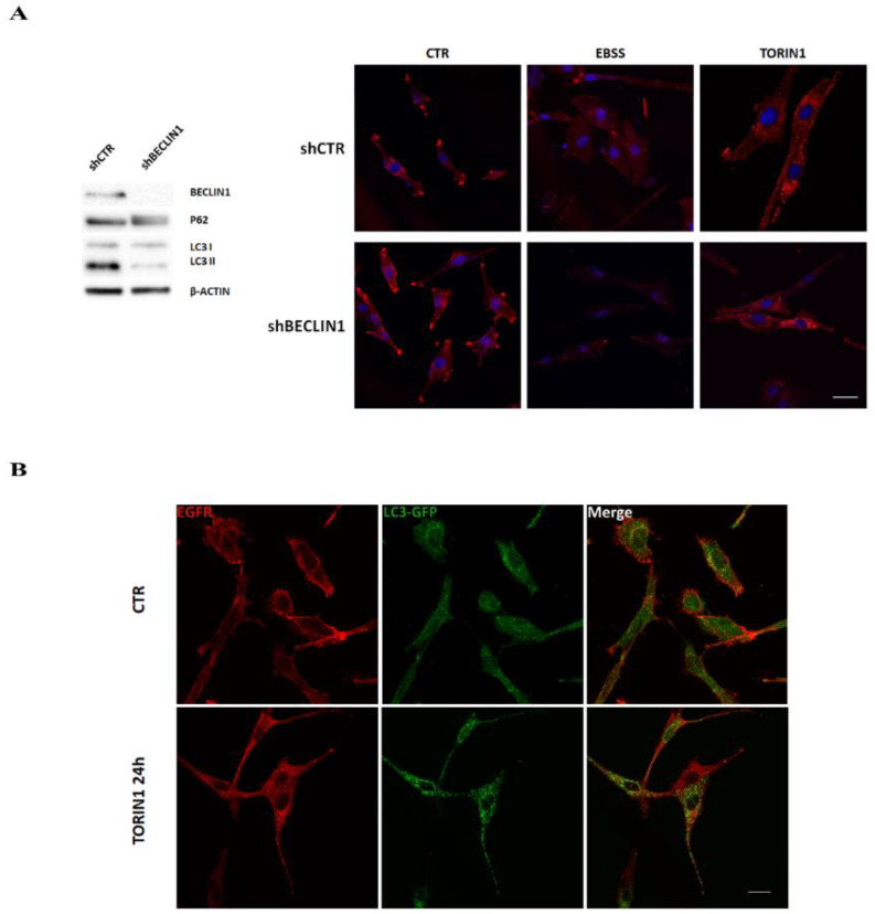
Figure 2.
EGFR de-localisation in GBM cells is independent of canonical autophagy. (A) shCTR and shBECLIN1 GL15 cells [23] were cultured in DMEM (CTR) or amino acid- and serum- free medium (EBSS) media or in DMEM in the presence of 250 nM Torin1 for 18 h and subjected to immunocytochemistry and confocal analysis for EGFR localisation (red) (right panels). Hoechst 33342 was used to stain nuclei (blue). Scale bar, 30 μM. Western blot analysis of P62 and LC3 I/II was also performed in basal conditions to check autophagy status (left panel). A specific antibody for BECLIN1 was used to check the silencing efficiency. β-ACTIN was used as loading control. The blot is representative of three independent experiments. (B) U87MG cells were transduced with GFP-LC3-expressing retrovirus as described in Material and Methods. Infected cells, cultured in DMEM alone (CTR) or in DMEM containing 250 nM Torin1 for 24 h, were subjected to immunocytochemistry and confocal analysis for EGFR (red) and autophagosomes (green) localisation. Colocalisation was excluded by calculating the Pearson’s correlation coefficient r (mean r CTR, 0.15 ± 0.02; Torin1, 0.2 ± 0.03). The images showing the merge of the two signals are shown in the right panels. Scale bar, 30 μM.

