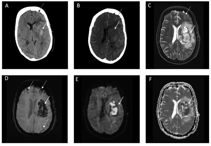
Figure 3.
Representative computed tomography (CT) and Magnetic Resonance Imaging (MRI) images of a patient with Coronavirus Disease 2019 with a deep parenchymal hemorrhage and subarachnoid hemorrhage (SAH). Legend: (A): Brain CT image with corresponding computed tomography (CTA) image indicative of no underlying vascular pathology (B) at admission. (C–F): Brain MRI image one day after hospitalization with T2w (C), susceptibility weighted imaging (SWI) (D), diffusion weighted imaging (DWI) (E), and corresponding apparent diffusion coefficient (ADC) imaging map (F). White arrow indicates acute intracerebral hemorrhage. White-black dotted arrow indicates acute SAH.

