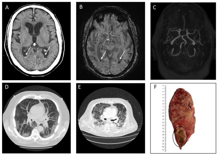
Figure 4.
Representative computed tomography (CT) and Magnetic Resonance Imaging (MRI) images of a patient with Coronavirus Disease 2019 with an intraventricular hemorrhage (IVH). Legend: (A,D,E): Brain CT image (A) with corresponding chest CT image 30 days after hospitalization (D). (B,C,E): Brain MRI image with susceptibility weighted imaging (SWI) (B) and Time of flight angiography (TOF) (C) with corresponding chest CT image (E) 32 days after hospitalization. (F): Longitudinal section of the right lung indicative of purulent pneumonia with abscess formation in the lower lobe. White arrow indicates acute intraventricular hemorrhage and black circle indicates abscess formation.

