Figure 3.
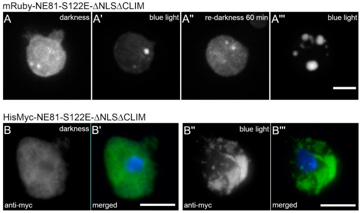
Blue light-induced assembly of NE81-S122EΔNLSΔCLIM is independent of the fusion tag. (A–A’’’) Selected time points of a spinning disk confocal live cell imaging with mRuby2-NE81-S122EΔNLSΔCLIM cells and excitation at 561 nm. Assembly was triggered by strong (40% AOTF transmission) exposure to 488 nm. (A) First recorded image after start of image acquisition; (A’) the same cell after 3 min strong (40% AOTF transmission) exposure to 488 nm; (A’’) the same cell after 60 min in the dark without 488 nm excitation; and (A’’’) the same cell after 3 min re-exposure to strong 488 nm laser light. Scale bar = 5 μm. (B–B’’’) Expansion microscopy of fixed HisMyc-NE81-S122EΔNLSΔCLIM cells stained with anti-Myc/anti-mouse-AlexaFluor 488 and Hoechst 33342. Merged images (B’,B’’’) show NE81-S122EΔNLSΔCLIM in green and chromatin in blue; (B,B’) without light; and, (B’’,B’’’) after blue light stimulation (band pass filter 450–490 nm). Expansion factors are 3.7 in (B) and 3.3 in (B’), respectively; scale bar = 5 μm (referring to the original size).

