Figure 5.
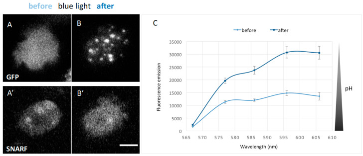
GFP-NE81-S122EΔNLSΔCLIM cells show a decrease in intracellular pH after excitation with blue light and form reversible cytosolic protein clusters. Live cells were loaded with the SNARF pH sensor. Blue light stimulation occurred for 30 s with a mercury halide lamp at a laser scanning confocal microscope (see methods): (A,A’) cells prior to blue light stimulation; and (B,B’) after blue light stimulation. The recombinant protein forms assemblies while SNARF fluorescence increases. Scale bar = 5 µm. (C) Emission spectra of SNARF-loaded GFP-NE81-S122EΔNLSΔCLIM cells (n = 8) excited at 561 nm and measured 3 min after blue light stimulation. The fluorescence emission of SNARF strongly increased between 565 and 610 nm.

