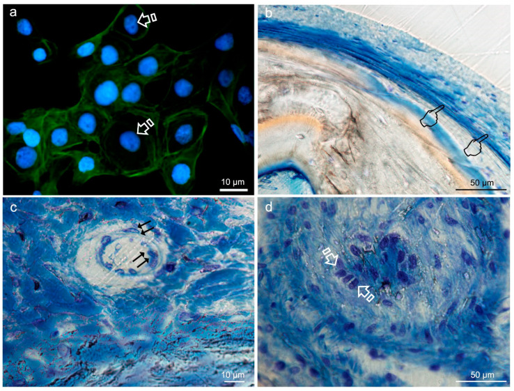
Figure 8.
Typical round-shaped images of the keratinocytes cytoskeleton (arrows) obtained by immunofluorescence, from Niculiţe et al. [26]. Copyright MDPI, 2018 (a). Bone histology obtained after using silica-loaded carboxilated doxycycline-doped membranes (b,c) and blank control (no membrane) (d), by coloration with toluidine blue to visualize the elongated fibroblasts (pointers in (b)), fusiform endothelial cells of blood vessels (double arrows in c) and cuboid osteoblastic cells (faced arrows in (d)), at 6 weeks of healing time in animal experimentation (Toledano et al./MAT2017-85999P).

