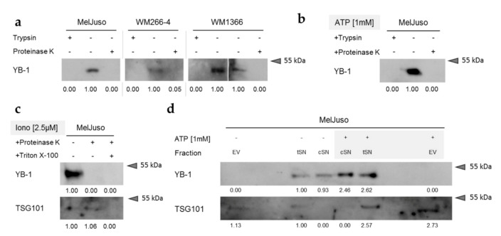
Figure 3.
YB-1 secreted from melanoma cells occurs mainly as a free protein. (a–c) Protease protection assay followed by Western blot analysis for YB-1 in serum-free cell culture supernatants. The cells remained untreated (a), were stimulated with ATP [1 mM] (b) or were stimulated with ionomycin [2.5 µM] (c). Culture supernatants were proteinase K (a–c) or trypsin (a,b) digested in the absence or presence of Triton X-100 (c) to disrupt vesicular membranes. The lumenal exosomal marker TSG101 serves as a positive control for intravesicular proteins. Relative band intensities are indicated relative to the untreated control supernatants and uncropped Western blots available in Figure S5. (d) Western blot analysis of MelJuso-conditioned serum-free cell culture supernatants before (total SN, tSN) and after (cleared SN, cSN) purification of extracellular vesicles (EV) by ultracentrifugation. The lumenal exosomal marker TSG101 serves as control for the successful purification or depletion of vesicles. Relative band intensities are indicated relative to the tSN of untreated cells and uncropped immunoblots available in Figure S6. Representative data of two independent experiments are shown.

