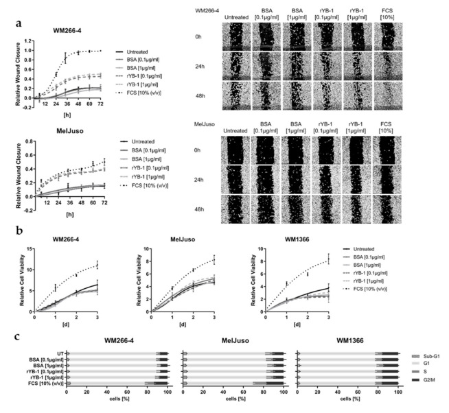
Figure 4.
Extracellular YB-1 stimulates wound healing capacity but not proliferation of melanoma cells. (a) Analysis of melanoma cell mediated wound closure after stimulation with recombinant YB-1 (0.1 µg/mL, 1 µg/mL), BSA as protein control (0.1 µg/mL, 1 µg/mL), or 10% FCS as positive control compared to the untreated (UT) cells (mean ± SD; N = 2 with n = 8; left panel). Representative pictures are shown after 0, 24, and 48 h (right panel). (b) Cell viability-based growth curves after stimulation with rYB-1 (0.1 µg/mL, 1 µg/mL), BSA (0.1 µg/mL, 1 µg/mL), or 10% FCS over 3 d. Representative data of two independent experiments are shown (mean ± SD, n = 6). (c) Flow cytometric cell cycle analysis of melanoma cells stimulated with rYB-1 (0.1 µg/mL, 1 µg/mL), BSA (0.1 µg/mL, 1 µg/mL), or 10% FCS for 24 h. Fractions of cells in sub-G1, G1, S, and G2/M phase were quantified (mean ± SD, N = 2 with n = 3).

