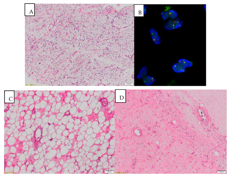
Figure 3.
(A) MLPS (myxoid liposarcoma) before treatment: paucicellular, monomorphic, myxoid tumor with plexiform vasculature and scattered signet ring lipoblasts (magnification 40×). (B) Rearrangement of the DDIT3 gene detected on the FISH analysis. (C) After treatment: striking lipomatous differentiation induced by RTH can be a predominant pattern after treatment (magnification 40×), (D) After treatment: diminished cellularity, stromal fibrosis, reduced vasculature with thickened blood vessel walls and tumor infiltrating macrophages (TAM). Area of necrosis in the right upper corner (magnification 40×).

