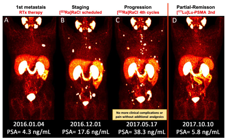
Figure 1.
(A–D) Sequential images (MIP) of [68Ga]Ga–PSMA PET/CT scan from a patient with metastatic prostate cancer. (A) He underwent local radiotherapy for single bone metastasis in the left 6th rib; (B) After approximately one year, he developed multiple painful bone metastases. He received 4 cycles of [223Ra]RaCl therapy. Pain reduced significantly and he no longer needed analgesic medication; (C) Nevertheless, the PSA level was apparently rising and the disease was progressing. The therapeutic plan was switched to [177Lu]Lu–PSMA. Afterwards, the response to therapy was clearly captured by [68Ga]Ga–PSMA PET/CT after 2 cycles of [177Lu]Lu–PSMA therapy, which was concomitant with the significant biochemical response (decrease in PSA level). [177Lu]Lu–PSMA: [177lutetium]Lu-prostate-specific membrane antigen; [223Ra]RaCl—[223radium]ra-dichloride; [68Ga]Ga–PSMA PET/CT—[68gallium] Ga-prostate-specific membrane antigen positron emission tomography/computed tomography; MIP—maximum intensity projection; PSA—prostate-specific antigen; RTx—radiotherapy.

