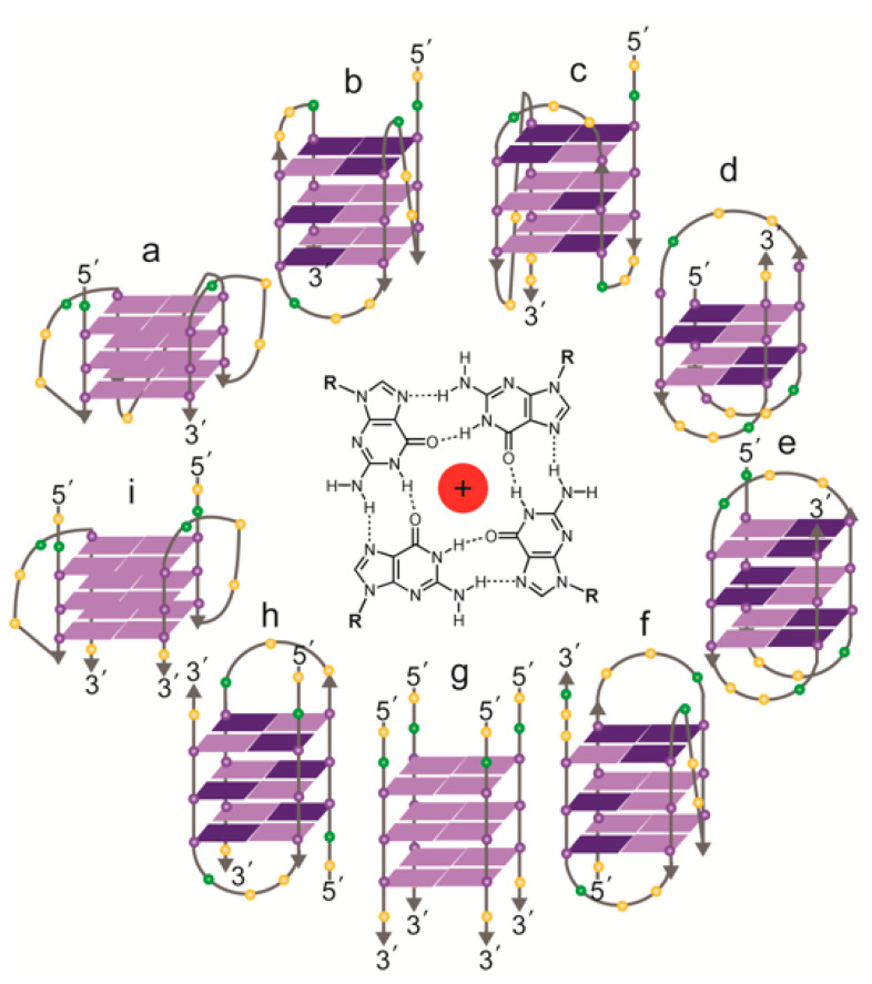
Figure 3.
Topologies of solved structures of human telomeric G-quadruplexes, either intramolecular (a–f) or intermolecular (g–i). Centre: a G-quartet, comprising four guanines, stabilized by a central cation. (a) Crystal structure of AG3(T2AG3)3 in K+ (parallel monomer) [84]; (b) NMR structure of TAG3(T2AG3)3 in K+ (hybrid form 1) [85]; (c) NMR structure of TAG3(T2AG3)3TT or TTAG3(T2AG3)3TT in K+ (hybrid form 2) [85,87]; (d) NMR structure of G3T2A(BrG)G2T(TAG3T)2 in K+ (antiparallel form 3) [86]; (e) NMR structure of AG3(T2AG3)3 in Na+ (antiparallel) [89]; (f) NMR structure of (T2AG3)3TTA(BrG)G2T2A in Na+ (antiparallel) [90]; (g) NMR structure of T2AG3T in K+ (parallel tetramer) [92]; (h) NMR structure of UAG3T(BrU)AG3T in K+ (antiparallel dimer) [93]; (i) crystal structure of (TAG3T)2 in K+ (parallel dimer) [84]; the same topology was seen in equilibrium with (h) by NMR with TAG3UTAG3T in K+ [93]. Guanines: purple spheres; thymines: yellow spheres; adenines: green spheres. Syn guanines shown in dark purple, anti guanines in light purple.

