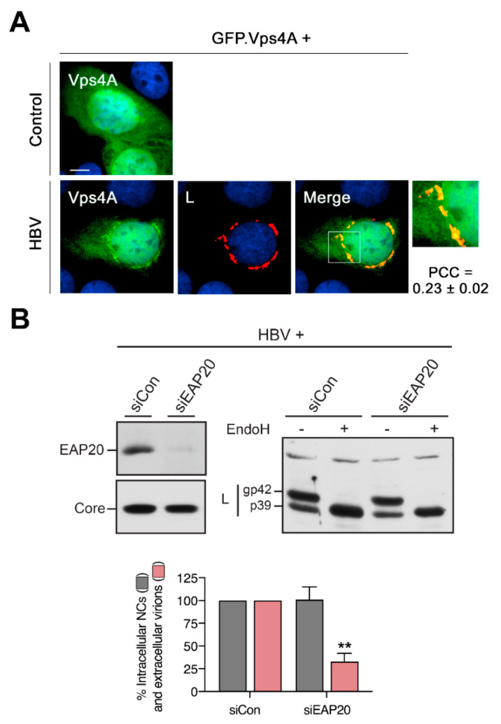
Figure 9.
Recruitment of ESCRT by the HBV-specific CS structure of L. (A) Cells were transfected with a GFP-tagged version of the Vps4A ATPase together with the control plasmid or the HBV replicon, followed by PFA fixation. The autofluorescence pattern of GFP.Vps4A is shown in green, anti-L labeling (57761, L) is in red and PCC values are indicated. (B) For RNAi of the ESCRT-II complex, cells were treated with EAP20-specific duplexes, transfected with the HBV* replicon and subjected to the virion production assay (n = 3, ± SD) exactly as described above. For a reference, a siCon treatment of cells was included. Lysates were probed by anti-EAP20 and anti-core (K46) immunoblotting. The same lysates were mock-treated or treated with EndoH prior to anti-L (K1350) WB. The L-specific p39 and gp42 forms are indicated. ** p < 0.01 compared to control.

