Figure 3.
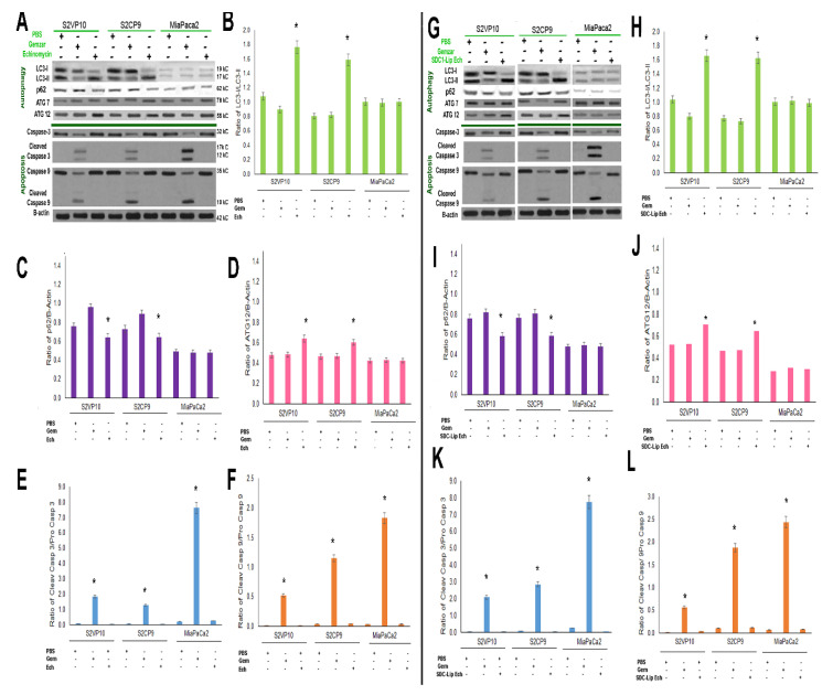
Mechanistic evaluation of SDC1-Lip echinomycin-induced autophagic (non-apoptotic) cell death using Western blot. As observed in Figure 2A,B, SDC1-Lip echinomycin induced cell death. To evaluate potential mechanisms of cell death, S2VP10, S2CP9, and Miapaca2 cells were treated with PBS, Gemzar, echinomycin alone, or SDC1-Lip echinomycin. Cell lysates were evaluated for signaling molecules important in autophagy (LC3-I, -II, p62, ATG7, and ATG12) and apoptosis (pro-caspase-3, cleaved caspase-3, pro-caspase-9, and cleaved caspase-9). (A–F) Echinomycin alone and (G–L) SDC1-Lip echinomycin treatment of S2VP10 and S2CP9 showed increases in the LC3-II/I ratio, indicating macroautophagy and lack of cleaved caspases-3 and -9, thus no apoptosis. Miapaca2 cells did not demonstrate activation of autophagy in response to either echinomycin or SDC1-Lip echinomycin. Control apoptosis treatment, Gemzar, resulted in some activation of apoptosis. Quantification comparisons are shown in B–F and H–L as well as Figure S6A–D. * p < 0.05.

