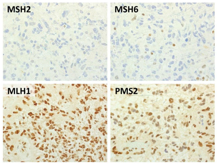
Figure 1.
Representative histological features of a MSH2/MSH6-negative glioblastoma. Immunohistochemical analysis highlighted negativity for MSH2 and MSH6 in neoplastic cells, with preserved expression of MLH1 and PMS2. Background inflammatory cells were used as positive internal controls. (Immunoperoxidase stain, original magnification 40×).

