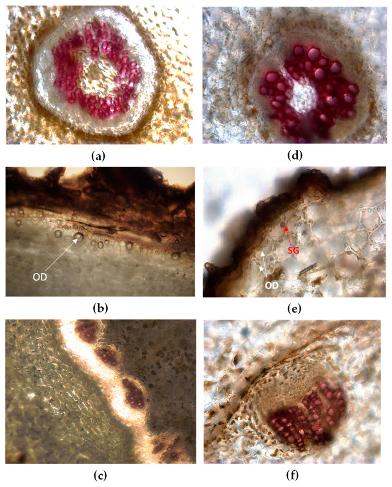
Figure 2.
Light microscopy view of root/rhizome transversal sections (TS) of Vo (a–c) and Vj (d–f) stained with phloroglucinol-HCl. Vo: (a) cortex and stele in older roots; (b) under the rhizome cork the outermost layers of parenchymatous cortex contain oil droplets (OD, arrow); (c) collateral vascular bundles circularly arranged in the rhizome. Vj: (d) cortex and stele in older roots; (e) starch grains (SG, arrow) and oil droplets (OD, arrows) are visible within parenchymatous cortex; (f) magnification of a singular collateral vascular bundle in the rhizome.

