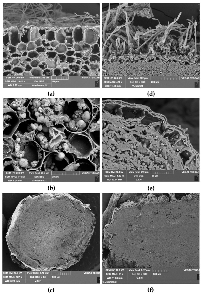
Figure 3.
Scanning electron microscopic view of TS of root/rhizome from Vo (a–c) and Vj (d–f): (a) epidermis with root hairs and the hypodermal layer of the cortex; (b) starch occurring as single or compound grains (2–6 components) within cortical parenchymatous cells; (c) TS of rhizome showing vascular bundles circularly arranged; (d) epidermis with many root hairs and parenchymatous cells filled with starch grains; (e) exoderm and parenchymatous cells filled with starch, generally occurring as single or compound grains with two components; (f) TS of rhizome with many vascular bundles surrounding the central pith.

