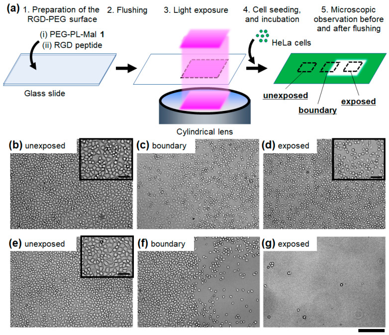
Figure 2.
Cell adhesion on the photocleavable RGD-PEG surface exposed to light before cell seeding. (a) Schematic illustration of the photo-responsive cell attachment. HeLa cells on the surface were observed at the (b,e) unexposed region, (c,f) the boundary region between the unexposed (left) and exposed regions (right), and (d,g) the exposed region. The bright-field microscopic images were obtained (b–d) before and (e–g) after flushing the non-adhered cells from the surface. Light dose: 4.0 J/cm2. Scale bars: 200 μm. (inset) Magnified images. Scale bars: 50 μm.

