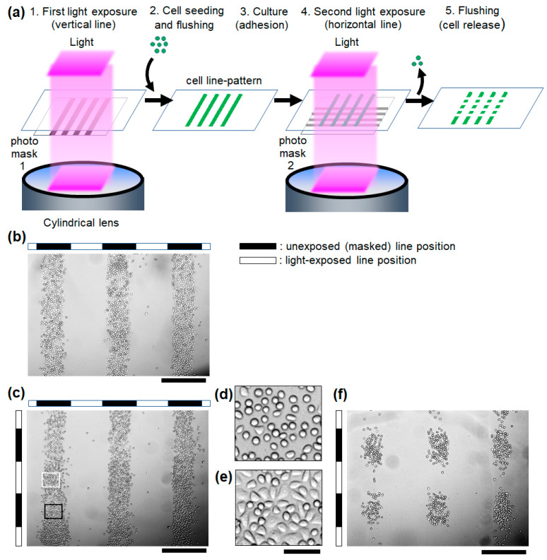
Figure 4.
Light-guided micropatterning and release of adherent cells on the photocleavable RGD-PEG surface. (a) Schematic illustration of the cell patterning and release. (b) The microscopic image of the vertical line-pattern of HeLa cells after cell adhesion to the first exposed surface. (c) Cells immediately after the second exposure of the horizontal line-pattern of light. Scale bars: 500 μm. Magnified image of the cells in the (d) light-exposed (white square in (c)) and (e) unexposed regions on the horizontal line-pattern of the second exposure (black square in (c)). Scale bars: 50 μm. (f) The remaining cell pattern after the second light exposure and rinsing. Scale bars: 500 μm.

