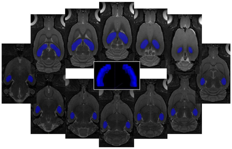
Figure 2.
In vivo MRI volumetry of the hippocampus of the rat brain. On 12 consecutive coronal T2-weighted MRI (resolution 0.137 × 0.137 × 0.5 mm3) is displayed representative regions of interest (ROIs) covering the hippocampus of the rat brain together with 3D visualization of target areas plotted by ITK-SNAP software (Version 3.4.0, US National Institutes of Health, Bethesda, MD, USA).

