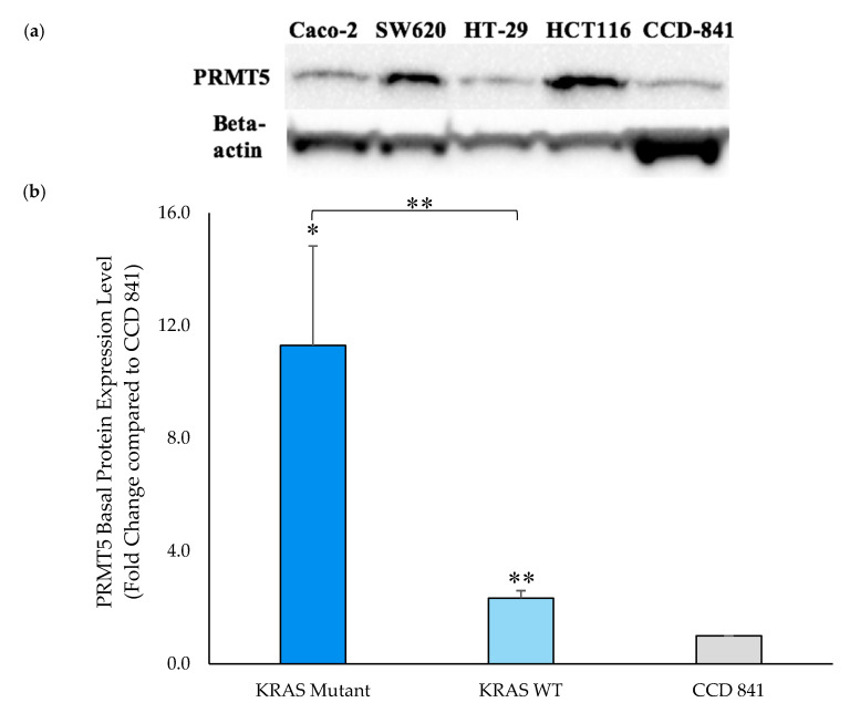
Figure 3.
PRMT5 is shown to be further overexpressed in the KRAS mutant CRC cells at the translational level by Western blot analysis. (a) Western blot assay results displaying PRMT5 and corresponding beta-actin bands; (b) Quantified Western blot assay results showing that PRMT5 protein is 4.8-Fold (p < 0.01) further overexpressed in the KRAS mutant CRC cells when compared to the KRAS WT CRC cells. Data are expressed as means + SD from two independent experiments using four CRC cell lines (2 KRAS mutant, 2 KRAS WT), as well as one normal colon cell line. The uncropped Western blot membranes can be seen in Figure S1. Data for the individual cell lines can be seen in Figures S2 and S3. * represents p < 0.05; ** represents p < 0.01.

