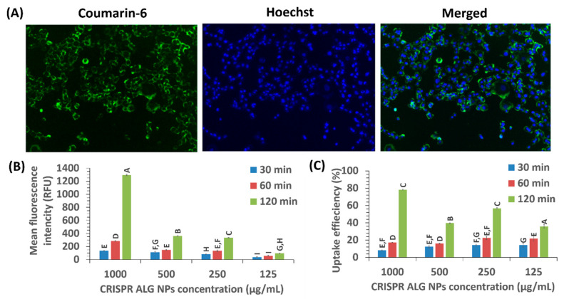
Figure 9.
(A) The cellular uptake of coumarin-6 loaded NPs in HepG2 cells at concentration 100 µg/mL at 37 °C for 2 h. The cell nuclei stained with Hoechst, and green color was the intrinsic fluorescence from courmarin-6. (B) The mean fluorescent intensity of NPs by HepG2 cells at various concentrations for 30, 60, 120 min. (C) Cellular uptake efficiency of NPs by HepG2 cells at various concentrations for 30, 60, 120 min. Letter denotes significance, whereby groups that do not share a letter are significantly different p < 0.05 (n = 3).

