Figure 7.
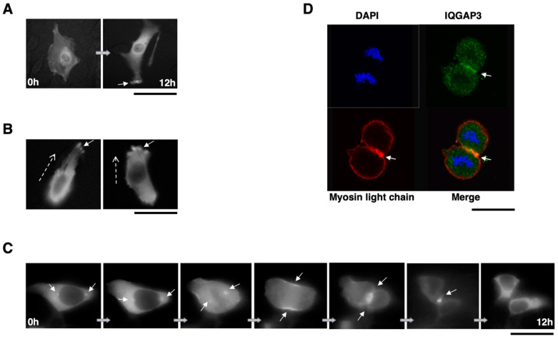
IQGAP3 is involved in cytokinesis. (A) Accumulation of IQGAP3 at the cell cortex area that subsequently underwent filopodia and lamellipodia formation in NIH3T3-IQGAP3 cells (arrow). (B) Accumulation of IQGAP3 at the leading edge of migrating cells (arrows). Dashed arrows; the direction of migration. (C) Representative images of subcellular localization of IQGAP3 during mitosis and cytokinesis in NIH3T3 cells expressing IQGAP3-GFP. IQGAP3 is localized at the spindle poles, contractile ring and cleavage furrow during cell division (arrows). (D) Co-localization of endogenous IQGAP3 and myosin light chain by immunocytochemical analysis in ST-4 cells. IQGAP3 and myosin light chain showed co-localization at the contractile ring during cytokinesis (arrow). Scale bars; 50 μm for (A)–(D).

