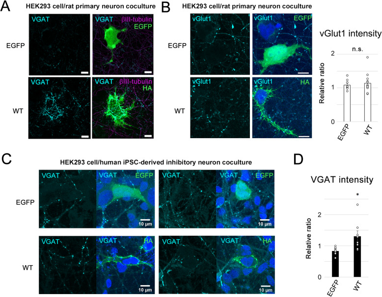
Fig. 3.
Synaptogenic activity of NL4X in rat primary neurons and human iPSC-derived inhibitory neurons. a Representative images of synaptogenic activity of WT NL4X expressed in HEK293 cells cocultured with rat primary neurons. Formation of inhibitory presynapse was visualized by immunostaining using anti-VGAT antibody. Quantification of the intensity of VGAT puncta was shown in Fig. 4a. Scale bar 10 μm. b Formation of excitatory presynapse was visualized by immunostaining using anti-vGlut1 antibody in the coculture of NL4X-expressing HEK293 cells and rat primary neurons. Quantification of vGlut1 puncta was shown at right (n = 10 (EGFP), 13 (WT), Welch’s t test; n.s., not significant). c Representative images of synaptogenic activity of WT NL4X expressed in HEK293 cells cocultured with human iPSC-derived inhibitory neurons. d Quantification of VGAT puncta in human iPSC-derived inhibitory neurons (n = 9 (EGFP), 8 (WT), *p < 0.05 vs WT by Welch’s t test)

