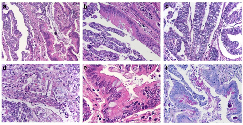
Figure 1.
Histological features of Endometrial Carcinoma showing intestinal-type features. (a) Endometrial carcinoma showing mixed endometrioid (right half) and intestinal-type mucinous (left half) features (H&E). (b) Single tumor gland showing concomitant endometrioid and intestinal-type differentiation (H&E). (c,d) Morphological details of the endometrioid component, composed by glands lined by columnar cells with scant cytoplasm (c), and foci of squamous differentiation (d) (H&E). (e) Morphological details of the intestinal-type component composed by enterocyte-like cells with apical brush border and goblet cells (H&E). (f) PAS-D istochemical stain highlighting the presence of intracytoplasmic mucin in the goblet cells of the intestinal-type tumor component (PAS-D stain).

