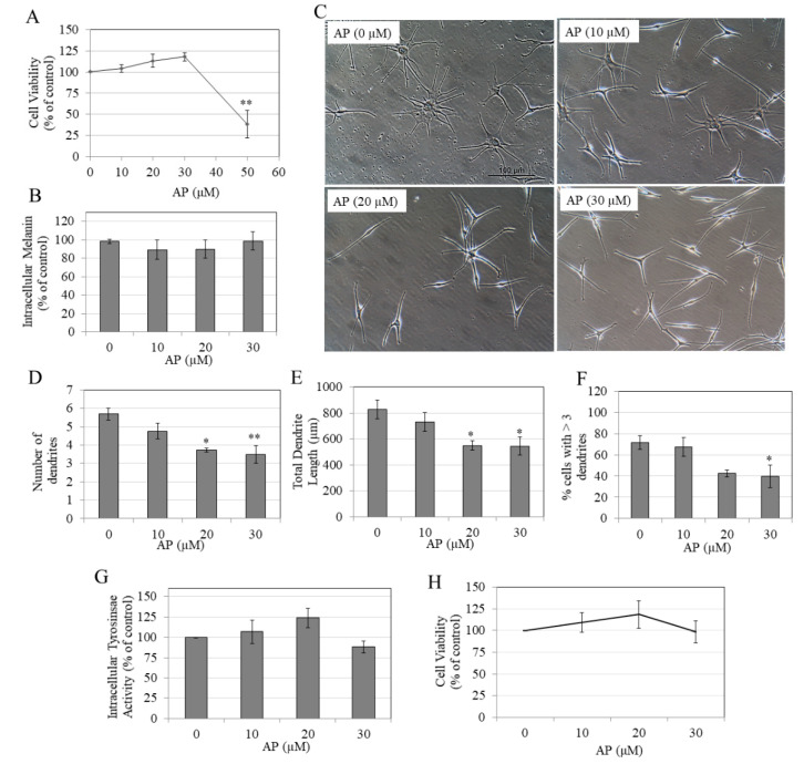
Figure 2.
(A) Viability of HEM-DP cells treated with AP at various doses for 5 d; ** p < 0.01 vs. Control group; one-way ANOVA with Dunnett’s test; (B) intracellular melanin levels; and (C) representative phase-contrast images of cells treated with AP (0–30 µM) taken at 20x objective magnification; melanocyte dendricity quantification by parameters: (D) Number of dendrites, (E) total dendrite length, and (F) % cells with >3 dendrites; a total of ~100 cells were counted for each treatment group; * p < 0.05 and ** p < 0.01 by one-way ANOVA with Dunnett’s test; (G) Intracellular tyrosinase activity in cultures of HEMn-DP cells treated with AP for 5 d; (H) viability of human keratinocytes (HaCaT) treated with AP at various doses for 5 d. All data are mean ± SEM of at least three independent experiments.

