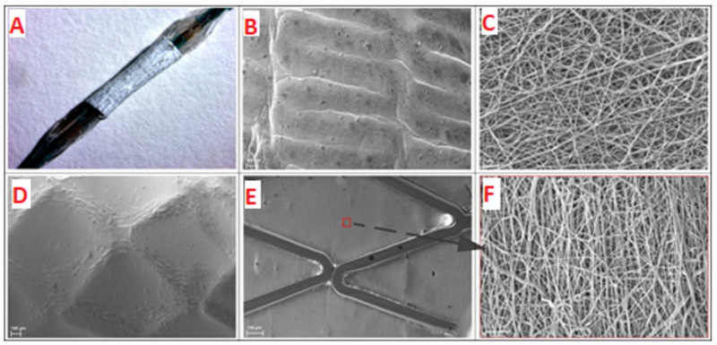
Figure 2.
A stent with the electrospun coating installed onto balloon catheter (A). Image obtained with SteREO Discovery V12 microscope (Carl Zeiss, Germany). SEM images of coating deformation after installation onto balloon catheter before (B,C) and after balloon expansion (D–F). Panels B,C,D show outer surface of electrospun coating; panels E and F show inner surface of electrospun coating (B,D,E panels—×149 magnification; C,F panels—×5000 magnification).

