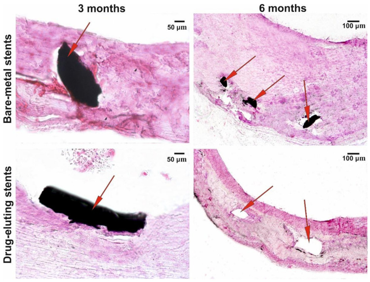
Figure 6.
Microscopy of the cross sections of implants at different observation points. Staining with hematoxylin and eosin; AxioLab A1 with the Zen 2 Blue Edition software package (Carl Zeiss, Oberkochen, Germany); magnifications, ×100 and ×400; arrows denote stent’s struts or the sites where the struts are located.

