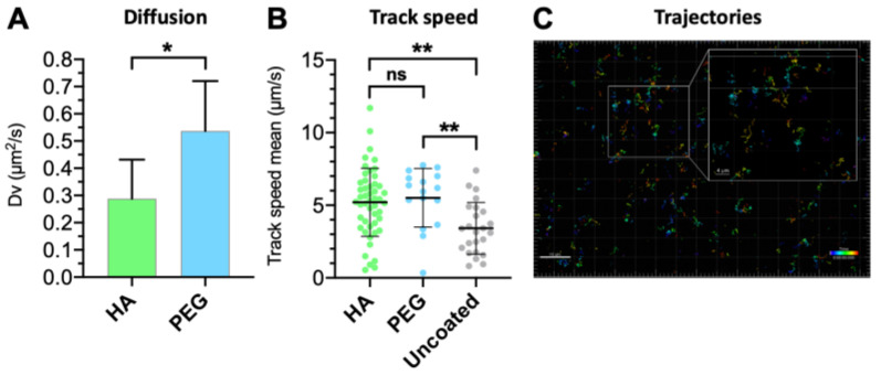
Figure 10.
Comparison between (A) vitreal diffusion of HA- and PEG-coated light-triggered liposomes, analyzed by unpaired t-test (* p < 0.05); (B) ensemble-average speed of HA-coated, PEGylated, and uncoated light-activated liposomes (τ = 1 s). Means with significant differences are shown as analyzed by ordinary one-way ANOVA with Tukey’s multiple comparison test (** p < 0.01). Error bars indicate SD. (C) Representative trajectories of the HA-coated liposome in the intact porcine vitreous. Inset shows the zoomed-in view of tracks obtained using the single-particle tracking technique.

