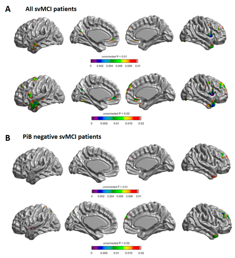
Figure 1.
Longitudinal change of cortical thickness in the white matter hyperintensity (WMH) progression group compared with the WMH regression group in all of the subcortical vascular mild cognitive impairment (svMCI) patients (A) and in PiB negative svMCI patients (B). Compared with the WMH progression group, the WMH regression group demonstrated more rapid cortical thinning in the coloured areas. Baseline age, sex, time, group, intracranial volume, Pittsburgh compound B standardised uptake value ratio, and the interaction between the groups and time were entered as fixed effects, and subject was entered as a random effect (uncorrected p < 0.01 in the upper row and p < 0.02 in the lower row).

