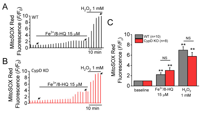
Figure 2.
Fe-induced reactive oxygen species (ROS) generation in ventricular myocytes isolated from WT and CypD KO mice. Increases in MitoSOX Red fluorescence were used as an indicator of mitochondrial superoxide production. MitoSOX Red fluorescence traces were recorded in a WT (A) and a CypD KO myocyte (B) treated with 15 µM Fe/8-HQ. The baseline value was normalized to 1, as indicated by the left-most arrow in each panel. The effect of Fe treatment was measured at the point indicated by the middle arrow. H2O2 (1 mM) was used as a positive control indicator. Summary data (C) were obtained from 10 WT and 8 CypD KO myocytes, ** p < 0.01 compared to baseline in WT and CypD KO group, respectively, by using paired t-test. NS: no significant difference was observed between WT and CypD KO groups by unpaired t-test.

