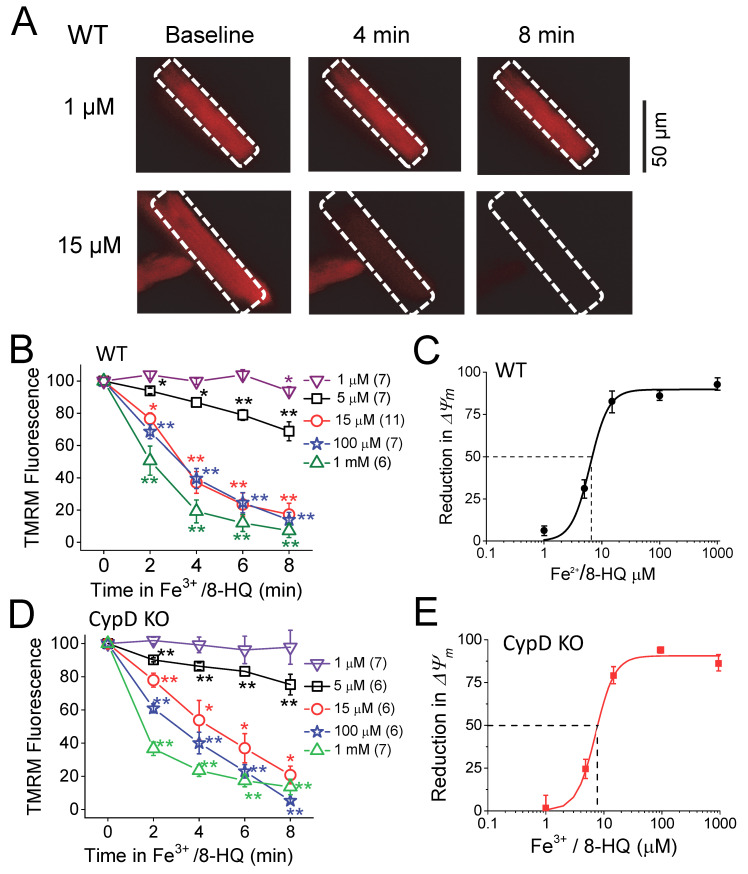
Figure 3.
Fe-induced depolarization of mitochondrial membrane potential (Δψm) in ventricular myocytes isolated from WT and CypD KO mice. (A) Representative snapshots of WT myocytes loaded with TMRM at baseline and 4 and 8 min after being treated with 1 and 15 µM Fe3+/8-HQ. (B) Summarized data showing a decrease in tetramethylrhodamine methyl ester (TMRM) fluorescence over time after Fe treatment in WT. The numbers of myocytes used in the measurement are indicated. (C) Percentile depolarization of ΔΨm at 8 min after Fe treatment in WT fit to Hill equation: ΔΨm = ΔΨm, max*[Fe]^nH/(EC50^ nH + [Fe]^nH). Half maximal effective dose (EC50) and Hill coefficient (nH) are 6.3 ± 0.3 µM and 2.7 ± 0.2, respectively, for WT. (D,E) In CypD KO myocytes, same as (B and C). EC50 and nH are 7.2 ± 0.4 and 2.7 ± 0.1 for CypD KO. * p < 0.05, ** p < 0.01 compared to the respective baseline value. Carbonyl cyanide p-(trifluoromethoxy) phenylhydrazone (FCCP, 30 µM) was used to completely dissipate the ΔΨm after the Fe treatment in each recording (not shown).

