Figure 5.
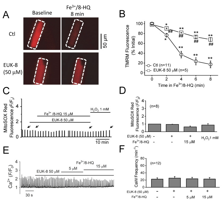
Attenuation of Fe-induced mitochondrial dysfunction and Ca waves by antioxidants. (A) Representative snapshots of WT myocytes loaded with TMRM at baseline and 8 min after being treated with 15 µM Fe3+/8-HQ in the absence or presence of 50 µM EUK-8. Arrows indicate the time points where fluorescence values were measured. (B) Summarized data showing a decrease in TMRM fluorescence over time after Fe treatment in WT (n = 11). * p < 0.05, ** p < 0.01, compared to baseline, respectively. ## p < 0.01 compared between control and EUK-8 groups (n = 5). (C) A representative MitoSOX Red fluorescence trace recorded in a WT treated myocyte with 15 µM Fe/8-HQ in the presence of EUK-8. (D) Summarized data of MitoSOX Red fluorescence recorded from 8 WT myocytes. (E) A representative CaW trace showing the effect of pretreatment with EUK-8 on the frequency of Fe-induced CaW formation in a WT ventricular myocyte. (F) Summarized data of the frequency of CaW formation recorded from 12 myocytes.

