Figure 3.
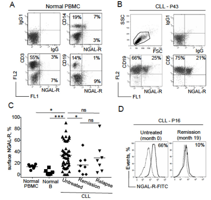
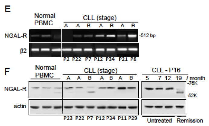
Expression profiles of NGAL receptor (NGAL-R) in PBMCs from healthy individuals, and CLL cells from patients before and after therapeutic treatment. (A) Representative cytograms of normal PBMCs stained with rabbit IgG-FITC/mIgG1-PE, NGAL-R-FITC/CD14-PE, NGAL-R-FITC/CD3-PE, NGAL-R-FITC/CD19-PE Abs. (B) Representative cytogram of CLL cells from one untreated patient, stained with rabbit IgG-FITC/mIgG1-PE, NGAL-R-FITC/CD19-PE and NGAL-R-FITC/CD5-PE Abs. (C) NGAL-R surface levels were determined, (with rabbit IgG-FITC and NGAL-R-FITC) in PBMCs and purified CD19+ B cells from healthy individuals, and CLL cells from patients before treatment or at remission or relapse. The data are presented as mean ± SEM (normal PBMCs, n = 7 healthy donors; isolated B cells, n = 6 healthy donors; CLL cells, n = 47 untreated patients, n = 8 patients in remission and n = 7 relapsed patients). p values were calculated using a Mann–Whitney U-test; not significant (ns); * p < 0.05; *** p < 0.001. (D) Expression of surface NGAL-R in CLL cells from patient P16 before treatment and at remission; cytogram of CLL cells stained with rabbit IgG-FITC (broken line) or NGAL-R-FITC Abs at diagnostic (month 0) and at remission at month 19 (month 19). (E) Representative RT-PCR results for NGAL-R in PBMCs from three healthy individuals and CLL cells from seven untreated patients. (F) Representative Western blot (reducing conditions) results for NGAL-R protein in PBMCs from three healthy individuals, CLL cells from seven untreated patients, and CLL cells of patient P16 before treatment (month 5, 7, and 12) and at remission (month 19).


